Image of the month (May 2006)

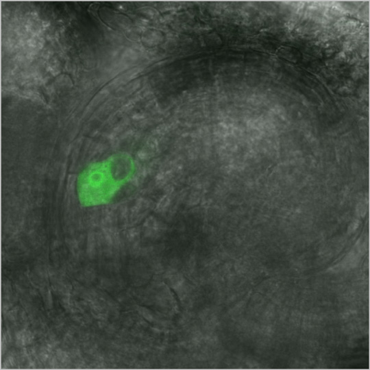
This is a confocal image of an Arabidopsis thaliana ovule. This image is from Fig. 3, Kasahara et al (2005) Plant Cell 17: 2981-2992.
In this paper, the authors report the functions of an A. thaliana gene, MYB98. To determine which cells within the female gametophyte expressed MYB98, they generated and analyzed transgenic A. thaliana plants containing ProMYB98:green fluorescent protein (GFP) constructs. Ovules from these plants were subjected to confocal microscopy analysis. This analysis showed that MYB98 is expressed only in the synergid cells until it is fertilized.
In this image, GFP expression can be seen in only one of the two synergid cells within an ovule. It is because the ovule is laid in such a way that the two synergids are on top of each other. The image was generated by merging bright field and fluroescent capture of the ovule; bright field image acuqisition allowed viewing of the all other ovule cells (which do not express GFP) possible.
Synergid cell specific GFP expression, synergid structure revelation and image acquisition with a perfect focus made this to be our image of the month.
-----------------------------------------------------------------------------
Click here for previously featured image of the month.
|